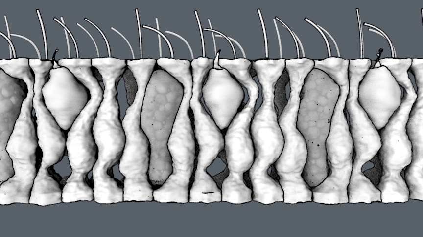
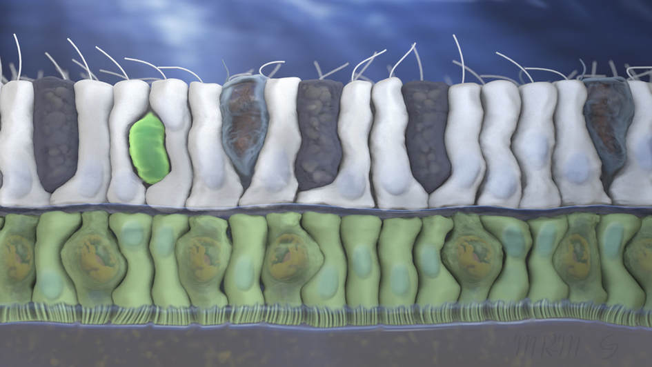
MEGAN RIEL-MEHAN
  • Home
  • Science Visualization
  • Research
    • Initial findings
    • Image processing
  • Civic Tech
  • Fine Art
  • News
  • Methods and Notes
  • CV and Resume

Visualizing the 
inner life of
coral

Coral Cell Biology


This was a joint project between our lab, the Birch Aquarium in San Diego and Martin Tresguerres Lab's at Scripps Institute of Oceanography on how coral cope with changing ocean chemistry. ​
Tresguerres Lab
Picture

Coral Nematocyte

Coral polyps have stinging cells similar to those in anemones and jelly fish. They consist of an inverted hollow tube that shoots out after the cell is triggered and barbs unfold to anchor the cell to the target. 
  Microscopy   video
Picture

Mucocytes

Coral polyps excrete mucous to trap food and cilia pump to move the mucus layer towards the polyp's mouth. 

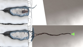
Early versions and drafts 

Here are some of the early work that lead to the final clips show above. 
Picture
Download CV
Contact me:
  • Home
  • Science Visualization
  • Research
    • Initial findings
    • Image processing
  • Civic Tech
  • Fine Art
  • News
  • Methods and Notes
  • CV and Resume