|
Traditional illustration techniques and computational approaches to visualize and analyze state of the art microscopy data
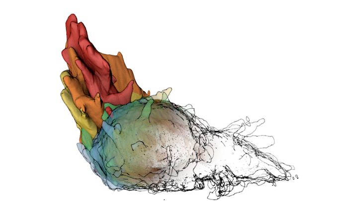
Megan Riel-Mehan The recently developed lattice light sheet microscope (LLSm) offers a unique combination of high spatial resolution, high speed and low phototoxicity. It is particularly well suited for visualizing fast moving, migrating cells such as white blood cells. However, the 3D time series data sets collected by the LLSm present many technical, visualization, and analysis challenges. Because this microscope is such a recent innovation, few existing analysis tools can visualize or process the data easily. In addition, there are limited visual conventions for exploring the data or communicating the results. The collaboration between the Mullins Cell Biology Lab (scientific visualization) and the Ferrin group (molecular viewing software) demonstrates how this shared project led to advances in our respective fields. Cell surfaces were imported into Cinema 4D then combined with more traditional illustration techniques of tonal shading and line to clarify surface topology and structural spatial relationships, among other complex analysis routines. The LLsm project exemplifies the many ways that visualization experts can help drive the scientific process and shows the benefits of engaging in collaboration early in the project timeline.
0 Comments
|


 RSS Feed